Engineering R&D: Putting contaminants in a new light
In the mid-1990s, microbiologists with the USDA’s Agricultural Research Service and a chemist from Iowa State University applied fluorescent spectroscopy to identify that indicator of fecal contamination on cattle carcasses. While others have applied light reflectance to address this food safety issue, the researchers were the first to develop a system that measures radiated light from a surface exposed to laser-induced light of a specific wavelength. By shining a blue light on the carcass, the still-light reactive pheophorbide emits a red light.
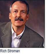
Sebastian, Fla.-based eMerge Interactive Inc. licensed the scientists’ discovery and prototype detection scanner and is about to roll out a commercial version. Food Engineering recently spoke with Richard Stroman, who heads the project.
An electrical engineer with a background in agricultural technology and image-processing systems, Stroman founded AgriVision Engineering Inc. in 1987, selling it seven years later to Key Technology. He served as Key’s director of engineering and helped develop several automated food inspection systems, including a cherry-pit detection system and a potato-strip defect removal system. Stroman joined eMerge Interactive in January 2000 and serves as vice president of intellectual property and advanced technology.
FE: How does fluorescent spectroscopy work?
Stroman: It uses the visual light spectrum, specifically the blue wavelengths, to illuminate and detect any organic matter on the carcass. Areas of contamination fluoresce at a different wavelength than the blue light, and that light can be filtered, amplified and processed by the system. A sophisticated and highly sensitive imaging system using multiple-wavelength cameras is required to detect ingesta or fecal contamination. The efficiency of fluorescent light is fairly low, so we have to amplify it considerably to make the system functional in a plant environment.
FE: What have been the engineering hurdles in bringing the detection system to market?
Stroman: The system uses the same core technology developed by Jacob Petrich of Iowa State University and Thomas Casey and Mark Rasmussen of USDA, but the device they designed was only a benchtop lab unit. eMerge received the rights to develop a commercial unit in late 1999.
Dealing with a fluorescent signal was a major challenge. When the light hits any microbial contamination, the fluorescence is converted to a different wavelength and reflected back at a very low level. The resulting signal has to be amplified significantly and discerned from everything else. There’s a lot of dynamic range in the carcass image, so the real challenge is applying the necessary subtraction techniques to highlight the microbes’ signal while filtering out the background signals.
FE: Why must the system be able to detect ingesta as well as fecal contamination?
Stroman: If the gut bursts during evisceration, it can spread ingesta—that is, food in the digestive track—throughout the internal area of the animal. The rumen can be a deadly host for the main three pathogens associated with beef: E. coli, Campylobacter and Salmonella.
FE: A side of beef has many irregular curves and crevices. Does this present an obstacle to detection?
Stroman: Because the carcass is split, the concave surfaces are exposed, and that makes illumination and detection easier. If it was whole, there would be a lot of shadowing, which would work against detection. With any surface that isn’t flat, you have to pay attention to the depth of field and make sure the three dimensions are in focus and evenly illuminated, and that was done.
FE: How quickly does the system generate operator feedback?
Stroman: Within a second, you know if the carcass needs additional cleaning or trimming. A second is an eternity in the image processing world. We have had to develop other systems that provide results within a fraction of a second. In this field, having a second to work with is a luxury.
If the managers so desire, they can choose to save images or compiled data generated for each side of beef for future reference. In effect, the system could serve as a critical control point in the HACCP plan.
FE: What are the unit’s physical dimensions?
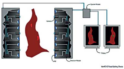
Stroman: For beef inspection, the system requires two 10-foot tall towers that are positioned on either side of the production line. The packer determines where they would be situated, but ideally we want to inspect carcasses after they’ve been split into two sides and while they are still on the hot side, before entering the cooler.
Three imaging modules are included in each tower, each of them responsible for illuminating about a three-foot area. They catch the leading and trailing edges of the carcass and provide a 360-degree view. Besides the detector modules, the tower includes lamps and an image-processing computer system. Data are relayed to a processor, which generates an image highlighting any areas of contamination. If anything is detected, the area is trimmed or vacuumed before entering the cooler.
FE: How long does the process take?
Stroman: Inspection is provided at full line speeds. The system inspects 600 to 700 half carcasses per hour on a four-foot center, or up to about 5,000 head of cattle per day. One unit could meet the needs of the largest beef packing plant.
FE: How might results from this system be used to adjust processes upstream?Stroman: If the operator notices certain patterns of contamination on the carcasses, that could be a clue to problems in a specific area. For example, if it looks like fecal contamination is being splattered, that might point to issues in the hide removal process. If the carcasses show signs of contamination from internal fecal material, that might point to problems in the way the animals are being eviscerated.
FE: Is fluorescent spectroscopy applicable to other kinds of livestock?
Stroman: We are a beef technology company, so that’s where we chose to start when developing the commercial system, but we have started feed-ration studies of swine and poultry to understand how their diets affect fluorescent light’s ability to see their fecal material. The cattle system is modular, with three vision processors in each tower to capture a full image. You don’t need three modules for swine, for example, so that might feature two processors, instead.
We’re developing a small hand-held device for use in inspecting small areas on beef. I think this technology has applications in food processing and infection control beyond livestock, as well.
FE: How much contamination must be present for detection to occur?
Stroman: Very low concentrations, down to the sub-micromolar level, well below what can be seen by the human eye.
FE: How does fluorescent spectroscopy fit with other food safety initiatives in packinghouses?
Stroman: A number of interventions out there today are somewhat effective, including carcass washing, organic acid rinses, steam pasteurization and steam vacuuming, but this is unique because it identifies specific areas of contamination. Some have suggested that processors simply wash the whole side of beef, but you might end up smearing contaminants and spreading them as effluent washes down the carcass.
FE: Prototype testing was completed a year ago. How soon will the system be commercially available?
Stroman: We’re well beyond the prototype stage and are in the final stages of developing the commercial system, which we hope to have in production late this year. We’ve been conducting various tests at Excel’s Schuyler, Neb., plant to fine-tune the camera system, image processing routines and user interface.
Looking for a reprint of this article?
From high-res PDFs to custom plaques, order your copy today!